


Mass Spectrometry

Mass Spectrometry is an analytical technique that is used for determining the molecular weight of various molecules and bio molecules. It provides structural information on proteins and conducts quantitative analysis with high sensitivity and specificity by sorting ionized analytes based on their mass-to-charge ratio. The mass spectrometric analysis or measurement of ionized analyte is carried out in the gas phase. Molecules are ionized and introduced in the electric field. In this electric field, they are sorted as per their mass-to-charge ratio. Mass spectrometry is used in many fields and applied to pure samples and complex mixtures.
In mass spectrometry, a sample in any state is ionized with a beam of electrons. Due to this ionization, some of the molecules in the samples break into fragments that are positively charged. In some cases there is no fragmentation, but only positively charged particles are formed. These positively charged ions are then separated according to their mass-to-charge ratio. Then, these ions are detected by an electron multiplier mechanism . Results are displayed as spectra of the signal intensity as a function of mass to charge ratio. Finally, atoms or molecules in the sample can be identified by their masses and correlate with known masses.
History of Mass spectrometry:
In 1886, Eugen Goldstein observed rays in gas discharges, and in 1899, he constructed a device with perpendicular electric and magnetic fields that separated the positive rays according to their charge-to-mass ratio (Q/m). Early spectrometry devices that measured the mass-to-charge ratio of ions were called mass spectrographs, which consisted of instruments that recorded a spectrum of mass values on a photographic plate. Modern techniques of mass spectrometry were devised by Arthur Jeffrey Dempster and F.W. Aston in 1918 and 1919, respectively.
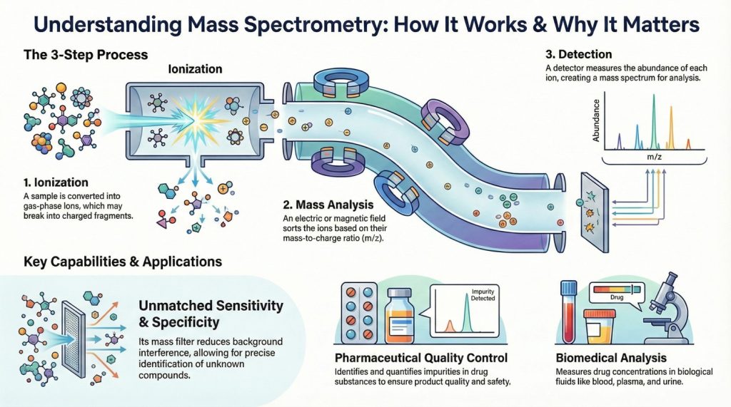
Different parts of Mass spectrometry:
A mass spectrometer has three distinct parts: an ion source, a mass analyzer, and a detector. The ionizer converts the sample portion into ions. Then, by the extraction mechanism, ions are removed from the samples. These ions then travel through the mass analyzer into the detector. The differences in the masses of the fragments allow the mass analyzer to differentiate the ions by their mass-to-charge ratio. The detector measures the value of an indicator quantity and thus provides data for calculating the abundances of each ion present. There are many types of mass analyzers, using either static or dynamic fields and magnetic or electric fields, but all operate according to the above differential equation. Each analyzer type has its strengths and weaknesses. Many mass spectrometers use two or more mass analyzers for tandem mass spectrometry (MS/MS).
Different types of ion sources are used as
Electron Ionization (EI):
A common method for generating ions from volatile compounds, it gives a high degree of fragmentation yielding detailed mass spectra. which can be used for the structural elucidation/characterization and identification of unknown compounds. However, EI is not suitable for coupling to HPLC, i.e. LC-MS, since at atmospheric pressure, the filaments used to generate electrons burn out rapidly. Thus, EI is coupled predominantly with GC, i.e. GC-MS, where the entire system is under high vacuum..
Electrospray Ionization (ESI):
Used for analyzing non-volatile and polar molecules, like peptides and proteins. The third one is Matrix-Assisted Laser Desorption/Ionization (MALDI), which is Used for analyzing large molecules, like proteins and peptides. Quadrapole, TOF, and Orbitrap are the commonly used mass analyzers. quadrupole is a very common type that is used to separate ions on the mass-to-charge ratio. In TOF separation is based on the time taken by the atom to travel a certain distance and allows the separation based on the mass. Orbitrap uses an electromagnetic field for the separation. It has a very high resolution compared to others.
Combination of Different separation Techniques with mass:
Different separation techniques are combined with the mass spectrometry to enhance mass resolving and mass determining capabilities. It is used in tandem with chromatographic and other separation techniques.
A common combination is Gas chromatography-mass spectrometry (GC/MS or GC-MS). In this technique, a gas chromatograph is used to separate different compounds. This stream of separated compounds is fed online into the ion source, a metallic filament to which voltage is applied. This filament emits electrons, which ionize the compounds. The ions can then further fragment, yielding predictable patterns. Intact ions and fragments pass into the mass spectrometer’s analyzer and are eventually detected. However, with the high temperatures used in the GC-MS injection port and oven, there is the possibility that the injected molecule may get degraded thermally, thus resulting in the measurement mass of degradation products instead of the actual molecule of interest.
Similarly to gas chromatography MS (GC-MS), liquid chromatography-mass spectrometry (LC/MS or LC-MS) separates compounds chromatographically before they are introduced to the ion source and mass spectrometer. It differs from GC-MS in that the mobile phase is liquid, usually a mixture of water and organic solvents, instead of gas. Most commonly, an electrospray ionization source is used in LC-MS. Capillary electrophoresis and ion mobility are the other technique that combines the liquid separation process of capillary electrophoresis with mass spectrometry. CE-MS is typically coupled to Electrospray ionization. Ion mobility spectrometry-mass spectrometry (IMS/MS or IMMS) is a technique where ions are first separated by drift time through some neutral gas under an applied electrical potential gradient before being introduced into a mass spectrometer.
Advantages and Disadvantages:
As an analytical tool of the new generation, it has its unique advantages and disadvantages over all other similar kinds of analytical tools. It has the greatest sensitivity among the instruments or analytical tools available. It is because it has a mass charge filter which reduces background interference and gives the highest specificity and sensitivity ti identify unknown or to confirm the presence of a compound. The disadvantage of the method is that it mostly fails to distinguish between optical and geometrical isomers and the positions of substituent in o-, m- and p- positions in an aromatic ring. Also, its scope is limited in identifying hydrocarbons that produce similar fragmented ions.
Applications of Mass Spectrometry: Mass spectrometry has both qualitative and quantitative uses. MS is now commonly used in analytical laboratories that study the physical, chemical, or biological properties of a great variety of compounds. Quantification can be relative or absolute. It is used to identify unknown compounds by analyzing the mass spectrum, It is used for the quantification of known compounds. It is also used for the structural analysis by understanding the fragmentation pattern of the molecule. It can be used for the determination of the ratio of isotope in an element. Mass spectrometry is used to analyze small as well as large molecules like metabolites and proteins. LC-MS helps identify and quantify impurities that may be present in drug substances or formulations, ensuring product quality and safety. LC-MS is used to measure drug concentrations in biological fluids (blood, plasma, urine) to assess drug absorption, distribution, metabolism, and excretion
FAQ:
Mass spectrometry (MS) identifies chemicals by ionizing them, separating the resulting ions based on their mass-to-charge ratio (m/z), and detecting their abundance, which is then displayed as a mass spectrum.
A mass spectrometer has three distinct parts: an ion source, a mass analyzer, and a detector. The ionizer converts the sample portion into ions..These ions then travel through the mass analyzer into the detector. The differences in the masses of the fragments allow the mass analyzer to differentiate the ions by their mass-to-charge ratio. The detector measures the value of an indicator quantity and thus provides data for calculating the abundances of each ion present.
References:
- Calabrese, Barbara. “Experimental Platforms for Extracting Biological Data: Mass Spectrometry, Microarray, next Generation Sequencing.” Elsevier EBooks, Elsevier BV, May 2018, pp. 126–29, https://doi.org/10.1016/b978-0-12-809633-8.20412-3.

